We perform ultrasound scans ranging from early pregnancy scans to 3D and 4D scans. All scans are performed by fully qualified and highly experienced sonographers.
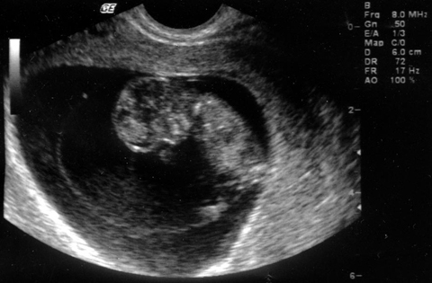
This scan is available for anyone who feels a bit worried or just wants to make sure that all is well with the pregnancy. It is also reassuring for women who have had some vaginal bleeding in early pregnancy (threatened miscarriage). These scans are usually done by means of an abdominal scanner. Sometimes the tummy scanner is inconclusive and an internal (vaginal) scan can give more information as to the health of the pregnancy. This scan can show the number embryos, a heartbeat, the size of the embryo and confirm the number of weeks of pregnancy.
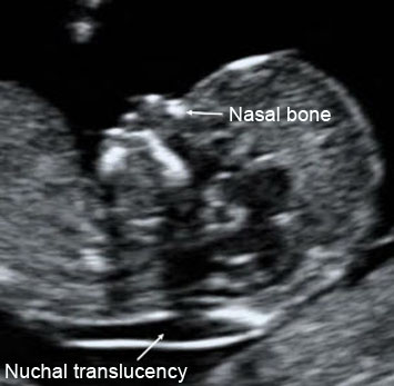
The Sonographer will measure the "nuchal fold" or "nuchal translucency". This is the fluid in the skin at the back of the baby's neck.. This fluid may be increased in babies with Down's syndrome or other genetic problem. If the nuchal translucency scan is abnormal, a referral to a Consultant in Fetal Medicine will be made and they will endeavour to see you on the day of your scan. You may be offered a definitive diagnostic test, amniocentesis, if the scan is abnormal.
A scan is not usually necessary at this stage of pregnancy but you might have one if you've had some bleeding or feel unwell. The baby will be measured on scan and the placenta and amniotic fluid will be assessed.
A scan at this stage can determine the sex of your baby. We also measure the baby at the scan. Our sexing scan are highly reliable but may be more difficult if the baby is in an unfavourable position or if the mother is overweight. We will not tell you the sex of the baby if we are not sure about it, but it is very unusual for us not to be able to do so.
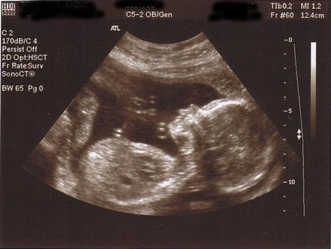
At the beginning of the scan, measurements will be made of the baby's head, brain, abdomen and legs. These will be plotted on a chart. We will then perform a detailed scan of the baby's organs to detect any abnormalities. This can never completely guarantee that all is well, but it gives very strong reassurance. We also check the placenta, amniotic fluid, and the umbilical cord. If you wish, you can find out the sex of the baby from this scan.
| Problem | What is the problem | Chances of being seen |
| Spina bifida | Open spinal cord | 90% |
| Anencephaly | Absence of top of head | 99% |
| Hydrocephalus | Excess fluid within the brain* | 60% |
| Major heart problem | 40% | |
| Diaphragmatic hernia | A defect in the muscle that separates the chest and the abdomen | 60% |
| Exomphalos/gastroschisis | A defect in the tummy wall | 90% |
| Major kidney problem | missing or abnormal kidney | 85% |
| Major limb abnormality | Missing bones or very short limbs | 15% |
| Cerebral palsy | Spasticity, weakness of limbs | not seen |
| Autism | not seen | |
| Down syndrome | May be associated with heart and bowel problems | about 40% |
| May be associated with heart and bowel problems about 40% | ||
If you are at risk of premature birth, it may be useful to have an internal scan (vaginal scan) to measure the length of your cervix. Sometimes, a short cervix at this time help to predict an early delivery.
This is also called a "fetal growth scan". During this scan, we measure the baby's head, tummy and thigh bone. These measurements will be used to calculate the baby's weight. We will also look at how the baby is moving, the amount of amniotic fluid around the baby and where the placenta is. We will also be able to look at the blood flow to the baby from the placenta through the umbilical cord using Doppler ultrasound.
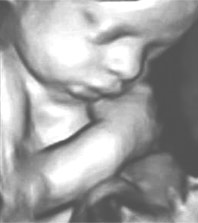
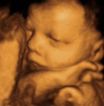
3D/4D scan are not usually need for medical reasons. Some people refer to them as "entertainment scans". If you are having a 3D/4D scan, we always do a wellbeing scan first before trying to view the face or other body parts. It may take some time to get a good view of the baby. If the baby is not in a favourable position, you may be asked to take a break and move around to see if the baby will also move.
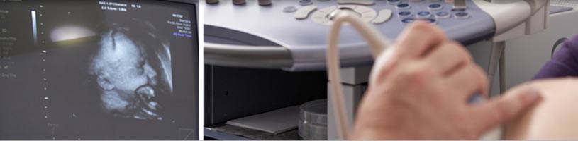
Our state-of-the-art scanning equipment provides the latest in ultrasound technology. The majority of gynaecology scans are performed using a vaginal probe (internal scan) to obtain the clearest views of the pelvis. This is done with an empty bladder. The uterus, cervix and both ovaries are visualised and measured. Any cysts or abnormalities are noted. A full report will be sent to your GP and/or Gynaecologist.